Nasal Turbinates
Causes, diagnosis and treatment options for chronic nasal obstruction caused by enlargement of the turbinates inside the nose.

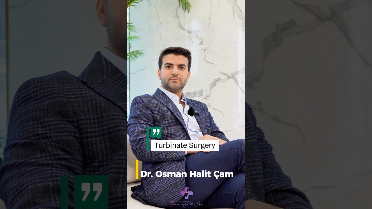
Doç. Dr. Osman Halit Çam
ENT & Head and Neck Surgery · Üsküdar, Istanbul
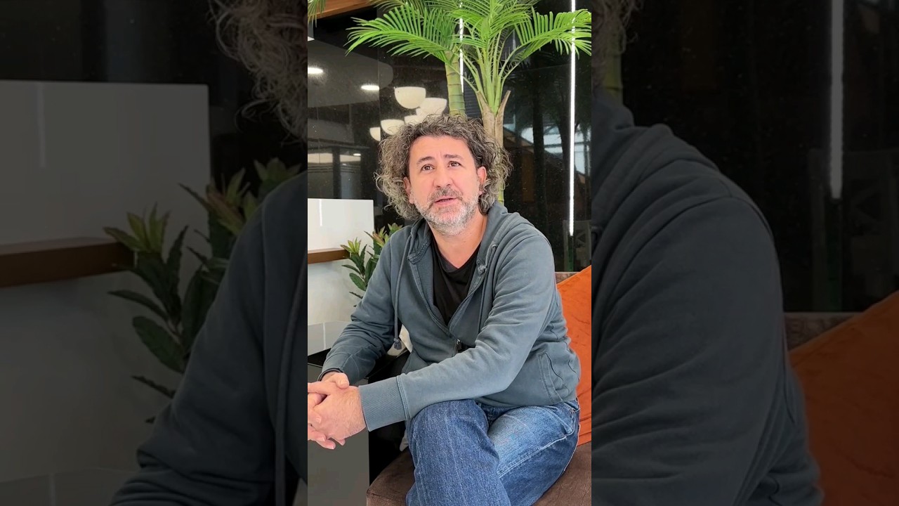
Nasal Turbinates — clinic video
{ AI · pre-assessment }
onlineLet's talk about your nose
AI responses are not a substitute for a medical diagnosis.
Konuyla ilgili kısa videolar
Tüm videolar →Doç. Dr. Çam'ın YouTube kanalından bu konuyla ilgili kısa açıklamalar. Toplam 2 video.
-
Burun Tıkanıklığında Konkalar — Nasıl Küçültülür?
Endolaser Turbinoplasti + Uyku Cerrahileri
The nasal turbinates are normal structures inside the nose that regulate airflow. They can enlarge and cause obstruction due to allergic rhinitis, chronic infection, decongestant spray overuse (rhinitis medicamentosa) or hormonal factors. Treatment ranges from medication to radiofrequency reduction; the goal is to shrink the turbinate while preserving its function.
- Type
- Medical + surgical
- Duration
- 20-40 minutes (surgical)
- Anesthesia
- Local / sedation
- Stay
- Same day
- Recovery
- 3-7 days
The nasal turbinates, known medically as conchae, are mucosa-covered structures on the side walls of the nasal cavity that regulate the airflow we breathe. Each side of the nose contains three pairs — inferior, middle and superior turbinates — and they warm, humidify and filter incoming air. The problem begins when a turbinate becomes permanently enlarged (hypertrophy); the nasal air passage narrows and the person feels constant nasal obstruction.
Summary: The turbinate is a functional structure inside the nose that warms and humidifies air. The inferior turbinate enlarges most often; the main causes are allergic rhinitis, chronic infection, rhinitis medicamentosa (decongestant spray overuse) and hormonal effects. The typical symptom is obstruction that alternates between sides and worsens at night. Treatment is stepwise: first medical care (allergy control, correct spray use), then radiofrequency reduction or turbinoplasty if needed. The core principle is that the turbinate is never removed completely, because excessive resection risks empty nose syndrome.
Table of Contents
- What Are the Nasal Turbinates?
- Why Do the Turbinates Enlarge?
- Symptoms: How Does Turbinate Enlargement Present?
- How Is the Diagnosis Made?
- Treatment Ladder
- Comparison of Treatment Approaches
- Septal Deviation and the Turbinates
- What Is Empty Nose Syndrome?
- Quality of Life and Sleep
- Turbinate Enlargement in Children
- For International Patients
- When Should an ENT Specialist Be Consulted?
- Frequently Asked Questions
- References
What Are the Nasal Turbinates?
The nasal turbinates are three pairs of structures — inferior, middle and superior — formed by a bony framework covered with mucosa along the side wall of the nose. The inferior turbinate is the largest and most richly vascularised, and is responsible for most obstruction complaints. The turbinates regulate and direct airflow within the nasal cavity.
In a healthy nose, the two sides swell and shrink in alternation within a rhythm called the nasal cycle, so it is normal for one side to feel slightly more blocked than the other during the day. The problem arises when a turbinate becomes permanently enlarged rather than cycling normally.
Why Do the Turbinates Enlarge?
Turbinate hypertrophy is usually due not to a single cause but to several overlapping factors. Allergic rhinitis, chronic infection, medication-related swelling and hormonal changes are the most frequent triggers, and identifying the correct one is decisive for whether treatment gives a lasting result.
1. Allergic rhinitis. Pollen, house dust mites, animal hair, mould and occupational allergens keep the mucosa in prolonged inflammation, so the turbinates appear swollen, pale and watery, with often seasonal symptoms.
2. Chronic infection and irritation. Recurrent sinusitis, postnasal drip, cigarette smoke, air pollution and dry, cold air can permanently thicken the mucosa over time.
3. Rhinitis medicamentosa (spray overuse). Long-term use of over-the-counter decongestant sprays causes persistent swelling that no longer opens even with the spray. Patients are often unaware of it; the solution is to stop the spray.
4. Structural causes and septal deviation. When the septum is deviated, the turbinate on the wider side enlarges over time to fill the space (compensatory hypertrophy), so the septum and turbinate are assessed together.
5. Hormonal and systemic causes. Pregnancy, hypothyroidism and certain blood-pressure medications can dilate the turbinate vessels and cause swelling.
Symptoms: How Does Turbinate Enlargement Present?
The most typical symptom of turbinate enlargement is nasal obstruction that alternates between sides during the day and worsens at night, with complete closure of the lower nostril when lying down. Seasonal symptoms suggest an allergic component, while year-round symptoms point more towards a structural or medication-related cause.
Common symptoms include:
- Constant or position-dependent nasal obstruction, marked at night
- Sleeping with the mouth open and waking with a dry mouth
- Snoring, sometimes with accompanying sleep apnoea
- Postnasal drip, frequent throat clearing and a reduced sense of smell
- Fullness around the forehead and eyes, headache and shortness of breath on exertion
- In children, constant mouth breathing, inattention and a decline in school performance
If these complaints do not resolve with medication, an evaluation is advised.
How Is the Diagnosis Made?
The diagnosis is largely made on examination, and advanced imaging is not needed in most cases. The aim is to distinguish whether the obstruction arises from the turbinate, the septum, the sinuses, or another structure such as a polyp. The evaluation consists of a few steps:
- History. Onset, seasonal pattern, spray use, previous nasal surgery, and allergy and sleep complaints are recorded.
- Anterior rhinoscopy and endoscopy. The nasal speculum shows the inferior turbinate and septum; a thin endoscope examines the posterior nose, middle turbinate, sinus openings and nasopharynx, revealing polyps or hidden deviations.
- Decongestant test. A short-acting decongestant temporarily shrinks the turbinate. If the obstruction fully resolves, the problem is usually mucosal swelling; if not, bone-related hypertrophy or another structural cause is more likely.
- Allergy testing and imaging (if required). A skin prick or blood test when allergy is suspected, and a paranasal sinus tomography if sinusitis or a polyp is suspected; routine tomography is not needed for the turbinates alone.
Treatment Ladder
The fundamental rule is this: the turbinate is never removed completely — it is reduced without impairing its function. Treatment proceeds in steps, and surgery is not always the first option. Medical treatment is tried first; if it is insufficient, reduction procedures or surgery come into consideration.
Medical (Conservative) Treatment
The first step is medical treatment, usually applied for 6-12 weeks; if it does not give enough relief, surgical options are considered. Correct spray technique and allergy control alone are often enough in many patients.
- Steroid nasal sprays (mometasone, fluticasone) — used regularly with correct technique, they shrink the mucosa over several weeks.
- Antihistamines — when an allergic component is present.
- Saline irrigation — for dryness and postnasal drip.
- Managing rhinitis medicamentosa — stopping the decongestant spray and switching to a steroid spray.
- Environmental measures — house dust control, humidity balance and avoiding cigarette smoke.
Radiofrequency Turbinate Reduction
Radiofrequency (RF) is the most frequently preferred minimally invasive method today. Under local anaesthesia, a thin probe delivers energy to the inner tissue of the turbinate while the outer mucosal surface is preserved. The office procedure is short and the patient goes home the same day; mild crusting occurs in the first few days, and the full effect appears within 4-6 weeks. Coblation and laser are other reduction methods used with a similar aim.
Surgical Reduction (Turbinoplasty)
In advanced turbinate hypertrophy, turbinoplasty (partial resection) or a submucosal microdebrider technique may be chosen. Part of the turbinate is reduced while the mucosa and functional tissue are preserved. If septal deviation is also present, the reduction can be performed in the same session as septoplasty. The choice of method is individualised according to turbinate size, the bone-to-mucosa ratio, allergic status and accompanying nasal problems.
The guiding principle is preservation: the turbinate is not removed completely, because excessive removal of functional tissue creates the risk of empty nose syndrome, discussed in the next section.
This content is for informational purposes only; diagnosis and treatment require an in-person physician examination. Surgical outcomes vary from person to person.
Comparison of Treatment Approaches
The table below summarises the three main approaches by who they suit and how durable they tend to be. The final decision rests with the physician based on examination findings.
| Approach | Who it suits | Anaesthesia | Durability |
|---|---|---|---|
| Medical treatment (spray, allergy control) | Mucosal swelling, allergic or early cases | None | Lasts while treatment continues; swelling may recur if allergy persists |
| Radiofrequency reduction | Moderate hypertrophy that does not open enough with medication | Local | Long-lasting; some cases may need repeating |
| Surgical reduction (turbinoplasty) | Advanced, bone-related hypertrophy; combined with septal surgery | Local / sedation | Close to permanent; functional tissue is preserved |
Septal Deviation and the Turbinates
Turbinate enlargement and a deviated septum (the nasal midline partition) frequently occur together, and patients often cannot tell them apart. When the septum is deviated to one side, the turbinate on the opposite side enlarges over time to fill the space (compensatory hypertrophy), so treating only one of the two is sometimes insufficient.
When the deviation is significant, septoplasty and turbinate reduction can be planned in the same session, with the evaluation weighing each structure’s contribution to the obstruction. The turbinate is also an important part of rhinoplasty planning, which addresses nasal aesthetics and function together.
What Is Empty Nose Syndrome?
Empty nose syndrome is a condition seen in patients from whom too much turbinate tissue was removed in the past. Although the nose appears open on examination, the person paradoxically experiences obstruction, dryness and a sensation of being unable to breathe, because the diminished tissue no longer regulates airflow normally. This is the most concrete reason why the turbinate should not be removed completely. The modern, preservation-focused approach reduces the turbinate while keeping functional mucosa and tissue, so that a balanced reduction keeps this risk to a minimum.
Quality of Life and Sleep
Turbinate enlargement is not in itself life-threatening, but because of its effect on sleep, oxygenation and daytime comfort it should not be neglected. Obstruction that worsens at night leads to sleeping with the mouth open, snoring and waking unrested. Where obstruction occurs together with snoring and sleep disturbance, a snoring and sleep evaluation is added to the plan. After turbinate adjustment, most patients notice improvement in night-time breathing, sleep quality and daytime concentration; if an allergic component persists, allergy management is continued after the procedure.
Turbinate Enlargement in Children
Isolated turbinate enlargement is less common in children than in adults, but it is frequently seen together with allergic rhinitis and adenoid enlargement. Constant mouth breathing, restless sleep, daytime inattention and a decline in school performance can be heralds of this picture. In a paediatric patient, the first-line approach is always conservative — steroid spray, allergy management, saline irrigation and, if necessary, assessment of the adenoids. Turbinate surgery is considered in children only rarely and in selected cases.
For International Patients
For patients travelling to Istanbul from abroad, the process usually begins with a remote consultation, where photographs, previous reports and the complaint are reviewed in advance so the examination and any procedure can be scheduled efficiently on arrival. A definitive plan is confirmed only after the in-person examination, since turbinate assessment depends on findings such as the decongestant test and endoscopy. Because most turbinate procedures are minimally invasive with rapid recovery, they often fit within a short stay, and the care team can help coordinate timing and follow-up in English.
When Should an ENT Specialist Be Consulted?
If symptoms do not resolve with medication or affect sleep and daily life, an ENT specialist should be consulted. Early evaluation can often resolve years of obstruction with a simple course of medication or a short office procedure. Assessment is advised for obstruction lasting more than 6 weeks that does not respond to medication, one-sided or progressive obstruction, an inability to breathe without a decongestant spray, obstruction that continues after previous septoplasty or rhinoplasty, or constant mouth breathing in a child.
Frequently Asked Questions
How can I shrink my turbinates without surgery? Regular, correct steroid-spray use shrinks swollen mucosa over several weeks, with allergy control and saline irrigation; if a decongestant spray habit exists, stopping it is the priority. Surgery is considered only if this falls short.
Does turbinate hypertrophy go away on its own? A temporary trigger such as a passing infection can subside, but persistent enlargement from allergy, structural factors or spray overuse usually does not resolve without treatment. It is managed medically first and reduced only if obstruction continues.
How risky is radiofrequency turbinate reduction? It is a minimally invasive office procedure under local anaesthesia and is generally well tolerated. Mild crusting and fullness may occur for a few days; individual response varies and results settle over several weeks.
Will a steroid nasal spray shrink the turbinates? Steroid sprays such as fluticasone or mometasone can shrink swollen mucosa, but the effect is gradual and depends on consistent, correctly aimed daily use over weeks. They work best when an allergic or inflammatory component is present.
Is the turbinate removed completely in surgery? No. Its function must be preserved, so the goal is reduction, not removal. Complete removal can lead to empty nose syndrome; surgery preserves functional tissue while reducing excess volume in a balanced way.
Can turbinate enlargement come back after treatment? If an underlying cause such as allergic rhinitis continues, the turbinate can swell again over time. Allergy management and correct spray use are therefore maintained afterwards, with regular ENT follow-up.
References
Frequently Asked Questions
How can I shrink my turbinates without surgery?
Regular, correct use of a steroid nasal spray shrinks swollen mucosa over several weeks, alongside allergy control and saline irrigation. If a decongestant spray habit is present, stopping it is the priority. Surgery is considered only if medical treatment is insufficient.
Does turbinate hypertrophy go away on its own?
When the trigger is temporary, such as a passing infection, swelling can subside. Persistent enlargement from allergy, structural causes or spray overuse usually does not resolve without treatment; it is managed medically first and reduced only if needed.
Is the turbinate removed completely?
No. Its function must be preserved; the goal is to reduce it. Complete removal can lead to empty nose syndrome. Surgery preserves the functional mucosa and tissue while reducing excess volume in a balanced way.
How risky is radiofrequency turbinate reduction?
It is a minimally invasive office procedure under local anaesthesia and is generally well tolerated. Mild crusting and fullness may occur for a few days. As with any procedure, individual response varies and results settle over several weeks.
Will a steroid nasal spray shrink turbinates?
Steroid sprays such as fluticasone or mometasone can shrink swollen turbinate mucosa, but the effect is gradual and depends on consistent, correctly aimed daily use over weeks. They work best when an allergic or inflammatory component is present.
When does the obstruction resolve after treatment?
Noticeable relief usually begins within 1-2 weeks, and the full result settles over about 4-6 weeks. During this period a "sometimes blocked, sometimes clear" sensation is normal as the mucosa reshapes.
Procedures often evaluated together
-
Septoplasty
Corrects the internal structure of the nasal septum to relieve chronic obstruction, with a noticeable effect on breathing, sleep and snoring; the external nose shape is unchanged.
-
Rhinoplasty
Rereading the architecture of the nose through function and proportion; aesthetic rhinoplasty where breathing and appearance are planned together.


Schedule a Consultation
Your information reaches Assoc. Prof. Dr. Çam's clinic. A response is made within 24 hours.